Computed Tomography – CT

Technique and Acquisition | Clinical Applications | Contrast Use and Protocoling | Role in Diagnostic Evaluation | Clinical Value

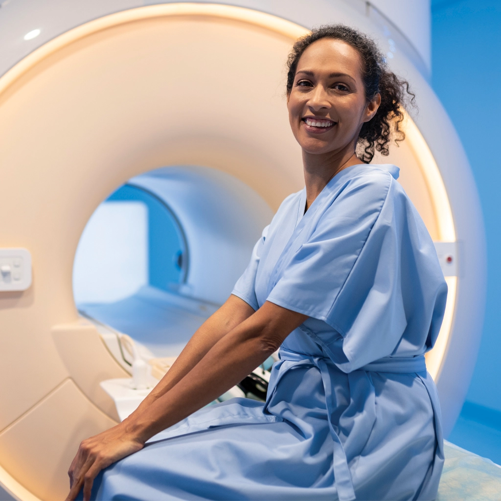
Computed tomography (CT) is used for cross-sectional imaging in both acute and non-acute clinical settings. It is often the first modality obtained when rapid diagnostic information is needed, particularly in emergency and inpatient environments where timely decision-making is critical. CT exams are performed across hospital-based and outpatient locations throughout Washington State and Alaska, allowing studies to be completed based on clinical urgency, patient location, and modality availability.

Computed Tomography - CT

CT Technique and Acquisition

CT imaging uses rotating X-ray systems to generate volumetric datasets that are reconstructed into multiple planes, including axial, coronal, and sagittal views. This approach allows for detailed visualization of internal structures within a short acquisition time. Imaging parameters are selected based on the clinical question and the area being evaluated, with attention to both image quality and efficiency.

In acute settings, protocols are designed to prioritize speed while maintaining diagnostic quality. In more complex scenarios, acquisition may be adjusted to provide additional detail of specific structures or areas of concern, depending on what needs to be clarified.

Clinical Applications

CT plays a central role in the evaluation of acute conditions such as trauma, intracranial hemorrhage, pulmonary embolism, and abdominal pain. In these settings, it provides rapid imaging, allowing providers to identify or exclude findings that may require immediate intervention. For this reason, CT is often the first cross-sectional study obtained in emergency imaging workflows.

Outside of acute care, CT is frequently used to evaluate infection, inflammatory conditions, and complex anatomical regions that require detailed cross-sectional imaging. In oncology, it supports the detection, staging, and follow-up of malignancies, allowing providers to monitor disease progression and treatment response over time.

CT is also used in vascular imaging, including evaluation of aortic pathology and thromboembolic disease, and in musculoskeletal imaging when fractures or complex bone anatomy require further characterization beyond X-ray.

Contrast Use and Protocoling

CT studies may be performed with or without contrast, depending on the clinical indication. Contrast-enhanced CT improves visualization of vascular structures, solid organs, and areas of suspected pathology. The decision to use contrast, along with the timing of image acquisition, is determined during protocoling to align with the diagnostic question.

When appropriate, imaging may be performed in multiple phases, including arterial, venous, or delayed phases. These are selected based on the structures being evaluated and the type of information needed from the study.

Role in Diagnostic Evaluation

CT is commonly obtained early in the diagnostic process to help define the next step in care. It is often performed after initial clinical evaluation or X-ray when additional detail is needed to guide management.

Findings on CT frequently determine whether further evaluation is required, including additional imaging, image-guided procedures, surgical consultation, or short-interval follow-up. In many cases, CT helps clarify the direction of care by narrowing the differential diagnosis.
Because of its speed and availability, CT may also be used at multiple points during care when reassessment is needed. This includes follow-up imaging to evaluate interval change, monitor known conditions, or assess treatment response.

Clinical Value

The strength of CT lies in its ability to provide rapid, detailed imaging that supports immediate clinical decision-making. It is widely used across a range of clinical scenarios because it balances speed, accessibility, and diagnostic accuracy.

In practice, CT is relied on when time matters and when a clear understanding of anatomy is needed to guide care.

Unlike traditional x-rays, CT images provide information about soft tissues inside the body and when combined with intravenous contrast also allow non-invasive evaluation of blood vessels.1


Contact us today for more information about CT scans, or click below to meet our radiologists.