Magnetic Resonance Imaging – MRI

MRI Technique and Image Detail | Clinical Applications | Contrast and Sequence Selection | Role in Diagnostic Evaluation | Clinical Value

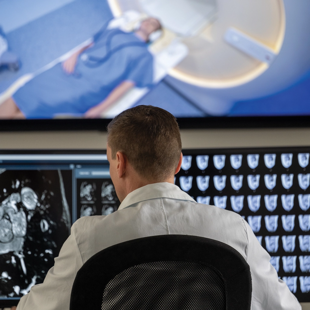
Magnetic resonance imaging (MRI) is used when detailed soft-tissue evaluation is required beyond what X-ray or CT can provide. It is commonly used to assess the brain, spine, joints, and other soft-tissue structures where subtle differences in anatomy or pathology need to be identified. MRI exams are performed across hospital-based and outpatient settings throughout Washington State and Alaska, supporting both scheduled imaging and follow-up evaluation after initial studies.

MRI - Magnetic Resonance Imaging

MRI Technique and Image Detail

MRI uses magnetic fields and radiofrequency signals to generate high-resolution images without ionizing radiation. Rather than relying on density differences alone, MRI produces images based on tissue characteristics, allowing for more precise differentiation between normal and abnormal structures.

Examinations are performed using a series of imaging sequences, each designed to highlight specific aspects of tissue composition, such as fluid content, structural integrity, or signal behavior. Images are acquired in multiple planes and can be adjusted to match the clinical question, providing flexibility in evaluating anatomy.

Studies are protocoled based on the clinical indication and the area of interest. Sequence selection is adjusted to ensure that the information obtained from the exam aligns with the diagnostic question, rather than relying on a fixed imaging approach.

Clinical Applications

MRI is most frequently obtained in neurologic and musculoskeletal imaging. In neurologic applications, it supports evaluation of conditions such as stroke, tumors, demyelinating disease, and degenerative changes of the brain and spine. Its sensitivity to subtle abnormalities makes it particularly useful when symptoms are not fully explained by prior imaging.

In musculoskeletal imaging, MRI provides a detailed assessment of ligaments, tendons, cartilage, and muscle. It is often performed when X-rays or CT scans do not fully account for a patient’s symptoms, or when a more detailed evaluation of soft tissue structures is needed. MRI can identify internal derangement and soft-tissue injury that may not be visible with other imaging techniques.

MRI is also used in abdominal and pelvic imaging when additional characterization is needed following CT or ultrasound. In these cases, it helps clarify findings that remain indeterminate after initial evaluation. It is also used in vascular imaging, including MR angiography, when evaluation of blood vessels is needed without iodinated contrast.

Contrast and Sequence Selection

MRI may be performed with or without contrast, depending on the clinical indication. Contrast is typically used to delineate lesions better, evaluate enhancement patterns, and assess disease activity, particularly in neurologic and oncologic imaging.

Sequence selection is a key component of MRI and is determined during protocoling. Different sequences provide complementary information about tissue characteristics, allowing for a more complete evaluation of the area being studied. The combination of sequences used in an exam is selected to match the clinical question and provide the most relevant diagnostic information.

Role in Diagnostic Evaluation

MRI is commonly obtained to evaluate further findings identified on prior imaging when additional detail is needed. In this role, it helps refine diagnosis by providing improved tissue characterization and a clearer understanding of the disease extent.

For example, an abnormality identified on CT may require an MRI to determine its composition or relationship to surrounding structures. Similarly, ultrasound findings may lead to an MRI when deeper or more complex anatomy needs to be assessed. MRI often provides the level of detail needed to determine whether intervention, surgical planning, or continued monitoring is appropriate.

MRI is also used for follow-up imaging, particularly to monitor disease progression or treatment response. Its ability to detect subtle changes over time makes it useful in conditions where small differences are clinically significant.

Clinical Value

The strength of MRI lies in its ability to provide detailed, high-contrast imaging that supports precise diagnosis. It is particularly valuable in situations where accurate tissue characterization is essential for guiding care.

In practice, MRI is relied upon when initial imaging raises questions that require further clarification, helping refine diagnosis and support clinical decision-making.

High-Field Open MRI – the same technology as a traditional MRI machine with a wider bore for the patient’s comfort. This wider bore allows for more space around the patient’s body while scanned. An Open MRI may help prevent claustrophobia and/or allow for larger patients to obtain an MRI scan.1


  1. Bellingham Advanced Medical Imaging – MRI Services

Contact us today for more information about MRI scans, or click below to meet our radiologists.