Mammography

Screening and Diagnostic Mammography | Imaging Technique | Clinical Applications | Role in Breast Imaging Workflow | Interpretation and Follow-Up | Clinical Value

Mammography is used to detect and evaluate breast disease and serves as a primary imaging tool for both routine screening and diagnostic assessment. It supports early identification of abnormalities that may not be clinically apparent. Mammography exams are performed across outpatient imaging centers and hospital-based settings throughout Washington State and Alaska, supporting both scheduled screening and follow-up diagnostic evaluation.

Mammography

Screening and Diagnostic Mammography

Mammography is performed in two main contexts: screening and diagnostic imaging. Screening mammography is obtained in patients without symptoms and is intended to detect early-stage breast cancer or other abnormalities before they become clinically evident. These exams are performed at regular intervals based on screening guidelines and individual risk factors.

Diagnostic mammography is performed when there is a specific clinical concern, such as a palpable finding, new symptoms, or an abnormality identified on a screening exam. In this setting, additional views are obtained to evaluate the area of concern further. Diagnostic exams are often performed in conjunction with a targeted ultrasound when additional characterization is needed.

Imaging Technique

Mammography uses low-dose X-ray imaging to evaluate breast tissue. Images are acquired in standardized views to support consistent evaluation and allow for comparison over time. Compression is applied during image acquisition to improve image quality and reduce motion artifacts, thereby providing clearer visualization of tissue structures.

Additional views or specialized techniques may be used during diagnostic evaluation to assess areas of concern better. The imaging approach may be adjusted during the exam based on the clinical indication and findings observed.

Clinical Applications

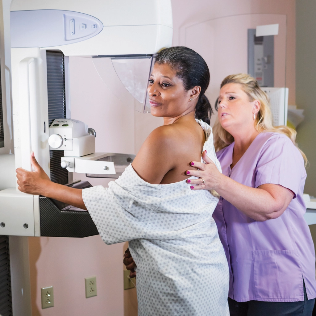
Mammography is used to detect a range of findings, including masses, calcifications, and architectural distortion. It plays a central role in the early detection of breast cancer, particularly when findings are subtle or not associated with symptoms.

In diagnostic settings, mammography is used to evaluate further abnormalities identified during screening or clinical examination. Findings are interpreted in the context of the patient’s history and prior imaging, when available. When appropriate, additional imaging or intervention may be recommended based on the results.

Mammography is often the starting point for breast imaging, with follow-up studies performed as needed to clarify findings or guide management.

Mammography also plays an important role in longitudinal breast imaging, where comparison with prior studies is essential. Subtle changes in tissue appearance over time can be as clinically significant as new findings, particularly in screening populations. For this reason, consistency in imaging technique and access to prior exams are important components of accurate interpretation. This longitudinal perspective allows radiologists to identify patterns of stability or progression that may influence management decisions.

Role in Breast Imaging Workflow

Mammography serves as the entry point for most breast imaging pathways. Screening exams identify patients who may require additional evaluation, while diagnostic exams help determine whether findings are benign, indeterminate, or require further intervention.

When additional imaging is needed, targeted ultrasound is commonly used to characterize findings further. In select cases, MRI may be used for additional evaluation, particularly in patients with more complex findings or higher risk profiles. If a suspicious finding is identified, image-guided biopsy may be recommended to obtain a definitive diagnosis.

This stepwise approach allows for structured evaluation, with each imaging stage contributing to clinical decision-making.

Interpretation and Follow-Up

Radiologists with experience in breast imaging interpret mammography studies. Findings are categorized and reported in a standardized format to guide follow-up recommendations. Interpretation takes into account prior imaging, when available, to assess for interval change.

Based on imaging findings, recommendations may include routine screening, short-interval follow-up, additional imaging, or biopsy. Communication of results is structured to support timely follow-up and appropriate management.

Clinical Value

The value of mammography lies in its role in early detection and structured evaluation of breast disease. By identifying abnormalities at an early stage, it supports earlier diagnosis and more effective treatment planning.

In practice, mammography functions as a central component of breast imaging, guiding further evaluation when needed and helping determine the appropriate next steps in care.

Tomosynthesis, also known as 3D mammography, is an advanced form of mammography that allows doctors to evaluate the breast tissue layer by layer. 3D mammography is more accurate and has been shown in studies to have a higher cancer detection rate as well as lower recall and false-positive rates.1


  1. Women’s Imaging Center – Mammography Services

Skagit Radiology is proud to partner with Skagit Regional Health to provide superior breast imaging at the Women’s Imaging Center.

Please contact the Women’s Imaging Center at 360-428-7275 to schedule a mammogram.